Quantcast
Channel: Species New to Science
Viewing all 10274 articles
Browse latest View live

[Herpetology • 2018] Hemidactylus malcolmsmithi • On the Status of Cyrtodactylus malcolmsmithi (Constable, 1949)

$
0
0

Hemidactylus malcolmsmithi  (Constable, 1949)

in Agarwal, Giri & Bauer, 2018.

Abstract
Cyrtodactylus malcolmsmithi was described by Constable in 1949 in the genus Gymnodactylus on the basis of its apparently undivided subdigital lamellae. The species has not been collected since and only finds mention in some checklists and new Cyrtodactylus descriptions. We recently examined the holotype and paratype of this enigmatic taxon and discovered that the subdigital lamellae are divided. The species is accordingly transferred to the genus Hemidactylus, within which it is a member of the Hemidactylus brookii complex and a valid speciesHemidactylus malcolmsmithi comb. nov. We assign recently sampled populations to this taxon and provide a diagnosis against congeners from the Indian subcontinent and a summary of characters for the species.

Keywords: Gekkonidae, HemidactylusHemidactylus brookii complex, Hemidactylus malcolmsmithi, India, South Asia

Figure 4. Hemidactylus malcolmsmithi in life (CES/11/050).
 Figure 2. View of left manus of Hemidactylus malcolmsmithi (left panel, CES/11/052 in life; right panel, holotype MCZ-R-3252).

Hemidactylus malcolmsmithi in life (CES/11/050).

Systematics
 Hemidactylus malcolmsmithi comb. nov. 
 Gymnodactylus malcolmsmithi Constable, 1949 
Cyrtodactylus malcolmsmithi Underwood, 1954
....

Natural History and Distribution.Hemidactylus malcolmsmithi is nocturnal and may be seen on the ground as well as low rocks, road cuttings, and buildings at night. The species is known from across the lowlands of Himachal and Jammu (up to about 1,500 m), and from a few specimens from Odisha and Rajasthan (Lajmi et al., 2016), though it is unclear what the native range of this species is, and which, if any, of these localities represent human translocations, with further sampling needed to determine its distributional range.

....

The status of the enigmatic taxon H. malcolmsmithi is finally resolved, through a combination of relatively recent field sampling, a careful examination of .140-year-old museum specimens, and recent publications on the H. brookii complex (Mahony, 2011; Lajmi et al. 2016). Constable initially did think he had a Hemidactylus before him, but the poor condition of the specimens and the opinions of two experts led him to place the species in Gymnodactylus. Interestingly, Khan (2010) opined that this species might be a misidentified specimen of H. brookii, and I.A. thought he might have this species when collecting Hemidactylus from around the Beas River basin (which we now know are in fact H. malcolmsmithi). However, the appearance of the lamellae in the types, which are longitudinally folded over themselves, had led previous researchers to erroneous conclusions.
....


 Ishan Agarwal, Varad B. Giri and Aaron M. Bauer. 2018. On the Status of Cyrtodactylus malcolmsmithi (Constable, 1949). Breviora. 557; 1-11.   DOI: 10.3099/MCZ41.1 


[PaleoIchthyology • 2018] Candelarhynchus padillai • A New Cretaceous Dercetid Fish (Neoteleostei: Aulopiformes) from the Turonian of Colombia

$
0
0

Candelarhynchus padillai 
Vernygora, Murray, Luque, Ruge & Fonseca, 2018


Abstract
The extinct neoteleost family Dercetidae includes elongate, long-jawed marine fishes that are known from the Late Cretaceous to Paleocene in deposits from the Levant, Europe, North Africa, England, Mexico and Brazil. The fossil record of the family in South America is very sparse and previously was restricted to outcrops in Brazil. Herein we describe a new dercetid fish from the Turonian of Colombia, †Candelarhynchus padillai gen. et sp. nov. A single articulated specimen is preserved in part and counterpart; the posterior part of the fish is missing. The specimen differs from other dercetid species by the following unique combination of morphological features: lack of scutes on the flanks of the body, presence of a single pair of transverse processes associated with the abdominal vertebrae, roofed posttemporal fossa, single row of small conical teeth on the dentary and maxilla, toothless premaxilla ornamented with pronounced longitudinal striations and protruding forward far beyond the anterior end of the dentary, and relatively large pectoral fins positioned high on the body. When included in a phylogenetic analysis of †Enchodontoidei, †C. padillai gen. et sp. nov. falls within a monophyletic family Dercetidae and is placed as sister taxon to the Late Cretaceous dercetid †Hastichthys from Israel, indicating faunal connections between the Eastern and Western Tethys. The new taxon provides novel insights into the distribution of dercetid fishes in the Western Tethys region during the early Late Cretaceous.

Keywords: Dercetidae, Enchodontoidei, Teleostei, San Rafael Formation, South America, palaeobiogeography


Oksana Vernygora, Alison M. Murray, Javier Luque, Mary Luz Parra Ruge & María Euridice Paramo Fonseca. 2018. A New Cretaceous Dercetid Fish (Neoteleostei: Aulopiformes) from the Turonian of Colombia. Journal of Systematic Palaeontology.  DOI: 10.1080/14772019.2017.1391884
ResearchGate.net/publication/321048665_A_new_Cretaceous_dercetid_fish_Neoteleostei_Aulopiformes_from_the_Turonian_of_Colombia
 twitter.com/JournalSystPal/status/930475709369344001

Ten-year-old boy helps paleontologists discover ancient fish species /phy.so/436700390 via @physorg_com
10-Year-Old Discovers Rare 'Lizard Fish' Fossil  on.natgeo.com/2EC3rXH via @NatGeo


[Botany • 2017] Alphonsea glandulosa • A New Species (Annonaceae) from Yunnan, China

$
0
0

Alphonsea glandulosa  Y.H. Tan & B. Xue

in Xue, Shao, Saunders & Tan, 2017. 

Abstract

Alphonsea glandulosa sp. nov. is described from Yunnan Province in south-west China. It is easily distinguished from all previously described Alphonsea species by the possession of glandular tissue at the base of the adaxial surface of the inner petals. Nectar was observed throughout the flowering period, including the pistillate phase and subsequent staminate phase. Small curculionid beetles were observed as floral visitors and are inferred to be effective pollinators since they carry pollen grains. A phylogenetic analysis was conducted to confirm the placement of this new species within Alphonsea and the evolution of the inner petal glands and specialized pollinator reward tissues throughout the family.

Fig 2. Flower and fruit morphology of Alphonsea glandulosa.
A, Branch, showing leaf-opposed inflorescence position, and lanceolate leaves. B, Abaxial view of the inflorescence, showing 5–6 carpels per flower. C, Adaxial view of the inflorescence, showing pubescent pedicels with one densely pubescent medial bract (arrowed). D, Fruit with subglobose monocarps. E, Single monocarp, dissected to show seed arrangement.
—Photos: Yun-hong Tan.

Alphonsea glandulosa is unusual in the genus, however, in having multiple flowers (often 5 to 9) in each inflorescence. Although most Alphonsea species have inflorescences with only 1–4 flowers, there are two species, A. philastreana (Pierre) Finet & Gagnep. and A. ventricosa (Roxb.) Hook. f. & Thomson, which have more than four flowers per inflorescence.
....

Alphonsea glandulosa Y.H. Tan & B. Xue, sp. nov.

Diagnosis: Alphonsea glandulosa is unique amongst Alphonsea species in having a nectar gland at the base of the adaxial surface of each inner petal. It is most similar to A. philastreana (Pierre) Finet & Gagnep., but differs in having a greater number of secondary veins on each side of the leaf, greyish to yellowish pubescent flower buds, longer pedicels, a greater number of carpels per flower, a smaller number of ovules per carpel, and globose to shallowly bilobed stigmas.

Etymology: The specific epithet reflects the presence of nectar glands at the base of the adaxial surface of each inner petal.


Bine Xue, Yun-Yun Shao, Richard M. K. Saunders and Yun-Hong Tan. 2017. Alphonsea glandulosa (Annonaceae), A New Species from Yunnan, China. PLoS ONE. 12(2); e0170107. DOI: 10.1371/journal.pone.0170107

A New Species of Alphonsea Found in Xishuangbanna
english.cas.cn/newsroom/research_news/201702/t20170204_173788.shtml

[Herpetology • 2018] Resurrection and Re-description of Plethodontohyla laevis (Boettger, 1913) and transfer of Rhombophryne alluaudi (Mocquard, 1901) to the Genus Plethodontohyla (Microhylidae, Cophylinae)

$
0
0

 [a-d] Individuals of Plethodontohyla laevis in life, illustrating the diversity of colour patterns from different sites of the species known distribution range.   Individuals of (A) Plethodontohyla alluaudi in dorsolateral and (inset) ventral view (ZSM 89/2004 , until now referred to as Pbipunctata; from Andohahela); (B) P. sp. Ca01 in dorsolateral view (ZCMV 555; from Ambatolahy)

in Bellati, Scherz, Megson, et al., 2018. 

Abstract
The systematics of the cophyline microhylid frog genera Plethodontohyla and Rhombophryne have long been intertwined, and their relationships have only recently started to become clear. While Rhombophryne has received a lot of recent taxonomic attention, Plethodontohyla has been largely neglected. Our study is a showcase of just how complex the taxonomic situation between these two genera is, and the care that must be taken to resolve taxonomic conundrums where old material, multiple genus transitions, and misattribution of new material obfuscate the picture. We assessed the identity of the historic names Dyscophus alluaudi (currently in the genus Rhombophryne), Phrynocara laeve and Plethodontohyla laevis tsianovohensis (both synonyms of Rhombophrynealluaudi) based on an integrative taxonomic approach harnessing genetics, external morphology, osteological data obtained via micro-Computed Tomography (micro-CT) and bioacoustics. We show that (1) the holotype of Dyscophus alluaudi is a member of the genus Plethodontohyla; (2) the Rhombophryne specimens from central Madagascar currently assigned to Rhombophryne alluaudi have no affinity with that species, and are instead an undescribed species; and (3) Phrynocara laeve and Dyscophus alluaudi are not synonymous, but represent closely related species, whereas Plethodontohyla laevis tsianovohensis is tentatively confirmed as synonym of D. alluaudi. We resurrect and re-describe Plethodontohyla laevis, and re-allocate and re-describe Plethodontohyla alluaudi on the basis of new and historic material.

Key Words: Amphibia, Anura, Phrynocara laeve, Plethodontohyla alluaudi, Madagascar, Integrative taxonomy


Figure 3. Individuals of Plethodontohyla laevis in life, illustrating the diversity of colour patterns from different sites of the species known distribution range:
(a) MRSN A6188 from Betampona in dorsolateral and (inset) ventral view; (b) MRSN A6181 from Betampona in dorsolateral view; (c) FAZC 13898 from Betampona in dorsolateral view (Photos by Gonçalo M. Rosa); (d) MRSN A6787 from Anivorano Est in dorsolateral and (inset) ventral view (Photo by Jasmin E. Randrianirina); (e) ZSM 189/2016 from Analalava-Foulpointe in dorsolateral view (Photo by Frank Glaw); (f) individual (not collected) from Ambodiriana in dorsolateral view (Photo by Lauric Reynes).

 Figure 4. Individuals of (a) Plethodontohyla alluaudi in dorsolateral and (inset) ventral view (ZSM 89/2004 , until now referred to as P. bipunctata; from Andohahela); (b) P. sp. Ca01 in dorsolateral view (ZCMV 555; from Ambatolahy); (c) P. brevipes in dorsolateral view (ZSM 649/2003; from Ranomafana); and (d) Rhombophryne sp. (formerly identified as R. alluaudi) in lateral view (ZFMK 52765 from Andasibe) (Photos by Frank Glaw and Miguel Vences).

 Adriana Bellati, Mark D. Scherz, Steven Megson, Sam Hyde Roberts, Franco Andreone, Gonçalo M. Rosa, Jean Noël, Jasmin E. Randrianirina, Mauro Fasola, Frank Glaw and Angelica Crottini. 2018. Resurrection and Re-description of Plethodontohyla laevis (Boettger, 1913) and transfer of Rhombophryne alluaudi (Mocquard, 1901) to the Genus Plethodontohyla (Amphibia, Microhylidae, Cophylinae). Zoosystematics and Evolution. 94(1); 109-135.   DOI: 10.3897/zse.94.14698


[Paleontology / Arachnida • 2018] Chimerarachne yingi • Cretaceous Arachnid illuminates Spider Origins

$
0
0

 Chimerarachne yingi  
Wang, Dunlop, Selden, Garwood, Shear, Müller & Lei, 2018


Abstract
Spiders (Araneae) are a hugely successful lineage with a long history. Details of their origins remain obscure, with little knowledge of their stem group and few insights into the sequence of character acquisition during spider evolution. Here, we describe Chimerarachne yingi gen. et sp. nov., a remarkable arachnid from the mid-Cretaceous (approximately 100 million years ago) Burmese amber of Myanmar, which documents a key transition stage in spider evolution. Like uraraneids, the two fossils available retain a segmented opisthosoma bearing a whip-like telson, but also preserve two traditional synapomorphies for Araneae: a male pedipalp modified for sperm transfer and well-defined spinnerets resembling those of modern mesothele spiders. This unique character combination resolves C. yingi within a clade including both Araneae and Uraraneida; however, its exact position relative to these orders is sensitive to different parameters of our phylogenetic analysis. Our new fossil most likely represents the earliest branch of the Araneae, and implies that there was a lineage of tailed spiders that presumably originated in the Palaeozoic and survived at least into the Cretaceous of Southeast Asia.


Bo Wang, Jason A. Dunlop, Paul A. Selden, Russell J. Garwood, William A. Shear, Patrick Müller and Xiaojie Lei. 2018. Cretaceous Arachnid Chimerarachne yingi gen. et sp. nov. illuminates Spider Origins. Nature Ecology & Evolution. DOI: 10.1038/s41559-017-0449-3


[Crustacea • 2018] Redescription of Parilia alcocki Wood-Mason, in Wood-Mason & Alcock, 1891 (Decapoda, Brachyura, Leucosiidae) from southeast India

$
0
0

Parilia alcocki   Wood-Mason, in Wood-Mason & Alcock, 1891

in Prema, Ravichandran & Ng, 2018.
 DOI:
 10.11646/zootaxa.4378.1.7 

Abstract

Parilia alcocki Wood-Mason, in Wood-Mason & Alcock, 1891, the type species of Parilia Wood-Mason, in Wood-Mason & Alcock, 1891, described from the Bay of Bengal, is one of the largest known leucosioid crabs, reaching carapace widths of over 50 mm. It is, however, not well known and its male gonopods have never been figured. A series of specimens of this species obtained from a fishing port in Tamil Nadu, India, allows for a comprehensive re-description and illustrations of the species. Its distinguishing characters from congeners are discussed.

Keywords: Crustacea, Deep-sea crab, Leucosioidea, Parilia alcocki, redescription, taxonomy


M. Prema, S. Ravichandran and Peter K. L. Ng. 2018. Redescription of Parilia alcocki Wood-Mason, in Wood-Mason & Alcock, 1891 (Decapoda, Brachyura, Leucosiidae) from southeast India. Zootaxa. 4378(1); 111–120.  DOI: 10.11646/zootaxa.4378.1.7


[Crustacea • 2018] Mayaweckelia troglomorpha • A New Subterranean Amphipod Species (Amphipoda, Hadziidae) from Yucatán State, México

$
0
0

Mayaweckelia troglomorpha Angyal

in Angyal, Solís, Magaña, Balázs & Simoes, 2018

Abstract
A detailed description of a new stygobiont species of the amphipod family Hadziidae, Mayaweckelia troglomorpha Angyal, sp. n. is given, based on material collected in four cenotes of Yucatán federal state, México. Morphology was studied under light microscopy and with scanning electron microscopy. Morphological description is complemented with mitochondrial cytochrome c oxidase subunit I (COI) sequences as barcodes, with affinities to the related taxa and with notes on the species’ ecology. Using COI Bayesian inference and genetic distance analyses, we show that the closest relative of the new species is M. cenoticola, forming a monophyletic group referring to the genus Mayaweckelia. Based on the available sequences, we also revealed that Mayaweckelia and Tuluweckelia are sister genera, standing close to the third Yucatán subterranean genus, Bahadzia. The data gathered on the habitat, distribution, abundance, and ecology will contribute to the conservation planning for M. troglomorpha Angyal, sp. n.

Keywords: cenote, description, endemic, Hadziidae, mitochondrial marker, morphology, SEM, sinkhole, subterranean


Figure 2. Mayaweckelia troglomorpha sp. n., living specimens.
Above: allotype ♀ collected in Cenote Kankirixché;
below: individual photographed in its natural habitat during research dive in Cenote Kanún (not collected).
Taxonomy
Order Amphipoda Latreille, 1816
Suborder Senticaudata Lowry & Myers, 2013

Family Hadziidae S. Karaman, 1943
Genus Mayaweckelia Holsinger, 1977

Mayaweckelia troglomorpha Angyal, sp. n.

Diagnosis: Medium-sized, eyeless hadziid with conspicuous troglomorphic traits. The first antenna almost twice as long as body and three times as long as the second antenna; gnathopod I propodus palm armed with distally notched spine teeth, carpus more than 1.5 times as long as corresponding propodus, merus as broad as but shorter than carpus, ventrally produced lobe with three long sensory setae; gnathopod II propodus twice as long as propodus I, palm armed with unnotched spine teeth, carpus slightly shorter than propodus on males. Dactylus, propodus. and carpus of pereopods VI-VII extremely long; therefore, pereopods VI and VII are 1.3 times as long as body length; epimeral plates I-III ventro-posterior corner tiny but distinct, ventral margin without robust setae, posterior margins concave; surfaces of uropods I-III pubescent; telson lobes each possess five-six robust setae and one-three slender setae on outer margin and six-seven robust setae on inner margin. Largest males and females both measured 10 mm.

Etymology: The name troglomorpha refers to the highly adaptive troglomorphic features of the new species, particularly the elongation of appendages, the increased number of sensory setae and papillae, and general appearance of fragility. Gender feminine.

Distribution and remarks on ecology: The new species is known from four cenotes in the state of Yucatán, covering a distribution distance of 52 km (distance between the farthest cenotes Dzonbakal and Xaan). All the individuals were found in fresh water habitat, in most cases far from the cenote entrances, deeper in the associated cave passages, where sunlight does not penetrate. Water temperature was between 26 and 27 °C. Specimens were collected between 20 and 33 meters depth; in cenote Kankirixché some individuals were observed below 45 meters depth. The new species was represented in all four localities with low abundance, though it proved to be more common and more abundant than M. cenoticola, of which a single specimen was found in only one (Ayun-Nah) of the 14 visited cenotes, during an underwater waste collecting activity, hidden in a plastic soft drink bottle. In the type locality and in cenotes Xaan and Kankirixché the new species co-occurred with the hadziid amphipod Tuluweckelia cernua. Other co-occurring stygobiont macro-crustaceans (in the four cenotes) were the mysid Antromysis cenotensis Creaser, 1936, the stygiomysid Stygiomysis cf. holthuisi (Gordon, 1958), the isopods Creaseriella anops (Creaser, 1936) and Yucatalana robustispina Botosaneanu & Iliffe, 1999, and the decapods Typhlatya mitchelli Hobbs & Hobbs, 1976, Typhlatya pearsei Creaser, 1936, and Creaseria morleyi (Creaser, 1936).

....

Conclusions: 
To date, only a small proportion of the cenotes and other aquatic hypogean ecosystems have been studied in Yucatán state in zoological aspect. Our expedition has led to the discovery of a new species of subterranean hadziids, which confirms that exploration and further studies of the region’s groundwater Crustacea diversity is necessary. Description of the new species was completed with comparative scanning electron microscopy, which was used for first time on Mayaweckelia. It proved to be a rather useful method for discovering, analysing, and illustrating barely visible diagnostic characters. As contributions to the future molecular genetic studies on Yucatán subterranean hadziids, COI sequences as barcodes of M. troglomorpha sp. n., M. cenoticola, and T. cernua are now publicly available in GenBank. The phylogenetic studies have shown that based on the available sequences, the closest relative of the new species is M. cenoticola. In accordance with the previous cladistic studies, Mayaweckelia and Tuluweckelia prove to be sister genera, closely related to Bahadzia, the third Yucatán subterranean Hadziidae genus. This knowledge may contribute to the species’ future conservation planning.

 Dorottya Angyal, Efraín Chávez Solís, Benjamín Magaña, Gergely Balázs and Nuno Simoes. 2018. Mayaweckelia troglomorpha, A New Subterranean Amphipod Species from Yucatán State, México (Amphipoda, Hadziidae). ZooKeys. 735; 1-25.  DOI: 10.3897/zookeys.735.21164

[Herpetology • 2018] Fritziana izecksohni • A New Bromeligenous Species of Fritziana Mello-Leitão, 1937 (Amphibia: Anura: Hemiphractidae) from High Elevations in the Serra Dos Órgãos, Rio de Janeiro, Brazil

$
0
0

Fritziana izecksohni 
 Folly, Hepp & Carvalho-e-Silva, 2018

Abstract
Fritziana is a genus of phytotelm-dwelling frogs that inhabits bamboo and bromeliads in the southern and southeastern portions of the Brazilian Atlantic Forest. Currently, five species are known, but the existence of undescribed species has been acknowledged. Here, we conducted a species-delimitation analysis of Fritziana using deoxyribonucleic acid sequences comprising two fragments of a mitochondrial gene (16S). The species delimitation analysis (with 16S) with Bayesian inference and maximum parsimony supported the recognition of a new species as an exclusive lineage, sister to all other Fritziana species. Bioacoustic and morphological evidence also support this finding. The new species occurs from 1166 to 2146 m above sea level and inhabits bromeliads from the National Park of Serra dos Órgãos, in the municipality of Teresópolis, state of Rio de Janeiro, Brazil. Compared with its congeners, the new species has nostrils narrowly separated, diameter of tympanum equal to or larger than diameter of disc on finger III, subarticular tubercles divided, and venter uniformly beige; advertisement call with notes composed of more than two pulses, and without the longer first pulse group.

Keywords: Atlantic rain forest, Bioacoustics, Molecular, Taxonomy


Manuella Folly, Fábio Hepp and Sergio P. Carvalho-e-Silva. 2018. A New Bromeligenous Species of Fritziana Mello-Leitão, 1937 (Amphibia: Anura: Hemiphractidae) from High Elevations in the Serra Dos Órgãos, Rio de Janeiro, Brazil. Herpetologica. 74(1); 58-72.  DOI:  10.1655/Herpetologica-D-16-00035


[Arachnida • 2018] Systematic Revision of the Giant Vinegaroons of the Mastigoproctus giganteus Complex (Thelyphonida: Thelyphonidae) of North America

$
0
0

AMastigoproctus vandevenderi, sp. nov., ♂. BM. giganteus (Lucas, 1835), ♀.
C
M. cinteotl, sp. nov., ♂. D. M. giganteus (Lucas, 1835), ♂.G. El Cielo, Municipio Gómez Farías, Tamaulipas, Mexico, habitat of M. cinteotl, sp. nov.  H. Puerto La Cruz, Municipio Yecora, Sonora, Mexico, habitat of M. vandevenderi sp. nov. 

Photographs: Griselda Montiel (C, G) 


Barrales-Alcalá, Francke & Prendini, 2018.
  Bulletin of the American Museum of Natural History. 418.
 DOI: 
10.1206/0003-0090-418.1.1 

 ABSTRACT
The North American vinegaroon, Mastigoproctus giganteus (Lucas, 1835), is demonstrated to comprise a complex of range-restricted species rather than a single widespread polymorphic species. Seven species are recognized based on morphological characters of the adult males, including the arrangement of spines on the prodorsal margin of the pedipalp trochanter, the position of the epistoma on the carapace, the presence of a stridulatory organ on opposing surfaces of the chelicerae and the pedipalp coxa, the presence of a patch of setae on sternite V, and the shape and macrosculpture of the retrolateral surface of the pedipalp femur. The two currently recognized subspecies are elevated to species: Mastigoproctus mexicanus(Butler, 1872), stat. nov., and Mastigoproctus scabrosus (Pocock, 1902), stat. nov.Mastigoproctus floridanus (Lönnberg, 1897) is revalidated from synonymy with M.giganteus. Redescriptions of M. giganteus and the other three species, based on both sexes, are provided, and three new species described:Mastigoproctus cinteotl, sp. nov., from Tamaulipas, Mexico; Mastigoproctus tohono, sp. nov., from Arizona and Sonora, Mexico; Mastigoproctus vandevenderi, sp. nov., from Sonora, Mexico. The present contribution raises the diversity of the Order Thelyphonida Latreille, 1804, in North America from one species to seven. Three species occur in the United States (one each in Arizona, Texas, and Florida), six species occur in Mexico, and two species occur in both countries.

Keywords: Arachnida, Uropygi, whip scorpion, biodiversity


FIG. 1. Species of Mastigoproctus Pocock, 1894, habitus in life (A–D) and representative habitats (E–H).
A. Mvandevenderi, sp. nov., ♂. B. M. giganteus (Lucas, 1835), ♀. C. M. cinteotl, sp. nov., ♂. D. M. giganteus (Lucas, 1835), ♂.
E. Cuernavaca, Municipio Cuernavaca, Morelos, Mexico, habitat of M. giganteus. F. Cascada de Atoyac, Municipio Atoyac, Veracruz, Mexico, habitat of M. scabrosus (Pocock, 1902), stat. nov. G. El Cielo, Municipio Gómez Farías, Tamaulipas, Mexico, habitat of M. cinteotl, sp. nov. H. Puerto La Cruz, Municipio Yecora, Sonora, Mexico, habitat of M. vandevenderi.
Photographs courtesy of Griselda Montiel (C, G) and Ricardo Paredes (E). 


Diego Barrales-Alcalá, Oscar F. Francke and Lorenzo Prendini. 2018. Systematic Revision of the Giant Vinegaroons of the Mastigoproctus giganteus Complex (Thelyphonida: Thelyphonidae) of North America. Bulletin of the American Museum of Natural History. 418; 1-62.  DOI: 10.1206/0003-0090-418.1.1


[Botany • 2016] Begonia titoevangelistae • A New Species (Begoniaceae, sect. Baryandra) from Catanduanes Island, the Philippines

$
0
0

Begonia titoevangelistae D. Tandang & R. Rubite

in Tandang, Rubite, Angeles & De Guzman, 2016. 

Abstract

Begonia titoevangelistae from Nahulugan Falls, Gigmoto, Catanduanes Island is described as a new species endemic to the Philippines. It resembles Begonia neopurpurea but is distinguished by the variegated leaves where the veins and midrib are dark green to almost black contrasting with light green interveins in the adaxial surface. In the abaxial surface, the veins are maroon contrasting with cream interveins, and its wide obliquely ovate leaf is elongated with shallowly undulate and minutely lobed leaf margin and long acuminate apex. Based on International Union for the Conservation of Nature red list criteria, B. titoevangelistae was assessed to be critically endangered, since it consists of one population with 300 individuals on a 100 m area around the waterfalls, which is being developed into a tourist spot.

Keywords: Begonia, Catanduanes, section Baryandra, critically endangered, Eudicots


Begonia titoevangelistae D. Tandang & R. Rubite

Etymology:—The species epithet is named after Dr. Luisito T. Evangelista the current Officer in-Charge— Curator I of the Botany Division, National Museum of the Philippines. Dr. Evangelista is fondly called “Sir Tito” by his colleagues thus the specific epithet titoevangelistae

Distribution and Habitat:—There was only one population discovered in June 28, 2015 at Nahulugan Falls, Gigmoto, Catanduanes Island. The plants grow on moist rocks under trees and shrubs near the waterfalls. 


Danilo N. Tandang, Rosario R. Rubite, Raul T. Angeles Jr. and Margaret C. De Guzman. 2016. Begonia titoevangelistae (sect. Baryandra, Begoniaceae) A New Species from Catanduanes Island, the Philippines. Phytotaxa. 282(4); 273–281. DOI:  10.11646/phytotaxa.282.4.4

[Paleontology • 2018] Mansourasaurus shahinae • New Egyptian Sauropod reveals Late Cretaceous Dinosaur dispersal between Europe and Africa

$
0
0

Mansourasaurus shahinae 
Sallam, Gorscak, O’Connor, El-Dawoudi, El-Sayed, Saber, Kora, Sertich, Seiffert & Lamanna, 2018

  DOI: 10.1038/s41559-017-0455-5 

Abstract
Prominent hypotheses advanced over the past two decades have sought to characterize the Late Cretaceous continental vertebrate palaeobiogeography of Gondwanan landmasses, but have proved difficult to test because terrestrial vertebrates from the final ~30 million years of the Mesozoic are extremely rare and fragmentary on continental Africa (including the then-conjoined Arabian Peninsula but excluding the island of Madagascar). Here we describe a new titanosaurian sauropod dinosaur, Mansourasaurus shahinae gen. et sp. nov., from the Upper Cretaceous (Campanian) Quseir Formation of the Dakhla Oasis of the Egyptian Western Desert. Represented by an associated partial skeleton that includes cranial elements, Mansourasaurus is the most completely preserved land-living vertebrate from the post-Cenomanian Cretaceous (~94–66 million years ago) of the African continent. Phylogenetic analyses demonstrate that Mansourasaurus is nested within a clade of penecontemporaneous titanosaurians from southern Europe and eastern Asia, thereby providing the first unambiguous evidence for a post-Cenomanian Cretaceous continental vertebrate clade that inhabited both Africa and Europe. The close relationship of Mansourasaurus to coeval Eurasian titanosaurians indicates that terrestrial vertebrate dispersal occurred between Eurasia and northern Africa after the tectonic separation of the latter from South America ~100 million years ago. These findings counter hypotheses that dinosaur faunas of the African mainland were completely isolated during the post-Cenomanian Cretaceous.


Life reconstruction of Mansourasaurus shahinae on a coastline in what is now the Western Desert of Egypt approximately 80 million years ago.
Illustration: Andrew McAfee


Hesham M. Sallam, Eric Gorscak, Patrick M. O’Connor, Iman A. El-Dawoudi, Sanaa El-Sayed, Sara Saber, Mahmoud A. Kora, Joseph J. W. Sertich, Erik R. Seiffert and Matthew C. Lamanna. 2018.  New Egyptian Sauropod reveals Late Cretaceous Dinosaur dispersal between Europe and Africa. Nature Ecology & Evolution.  DOI: 10.1038/s41559-017-0455-5
منصوراصورس   facebook.com/HeshamSallam

اكتشاف الديناصور المصري «منصوراصورس» https://www.scientificamerican.com/arabic/articles/news/discovery-of-the-egyptian-dinosaur-mansourasaurus/ via @forscience_ar


[Crustacea • 2018] Parallel Saltational Evolution of Ultrafast Movements in Snapping Shrimp Claws

$
0
0


Kaji, Anker, Wirkner & Palmer, 2018.

Highlights
• The evolutionary history of remarkable snapping claws in shrimp is reconstructed
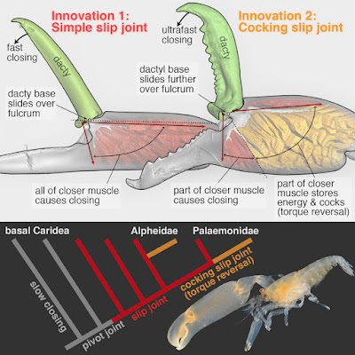
• Two novel claw-joint types—slip joints and torque-reversal joints—preceded snapping
• The transition “slip joint → torque-reversal joint → snapping” occurred in two families
• Subtle changes in joint form yielded dramatic changes in claw function (e.g., speed)

Summary
How do stunning functional innovations evolve from unspecialized progenitors? This puzzle is particularly acute for ultrafast movements of appendages in arthropods as diverse as shrimps, stomatopods, insects, and spiders. For example, the spectacular snapping claws of alpheid shrimps close so fast (∼0.5 ms) that jetted water creates a cavitation bubble and an immensely powerful snap upon bubble collapse. Such extreme movements depend on (1) an energy-storage mechanism (e.g., some kind of spring) and (2) a latching mechanism to release stored energy quickly. Clearly, rapid claw closure must have evolved before the ability to snap, but its evolutionary origins are unknown. Unearthing the functional mechanics of transitional stages is therefore essential to understand how such radical novel abilities arise. We reconstructed the evolutionary history of shrimp claw form and function by sampling 114 species from 19 families, including two unrelated families within which snapping evolved independently (Alpheidae and Palaemonidae). Our comparative analyses, using micro-computed tomography (microCT) and confocal imaging, high-speed video, and kinematic experiments with select 3D-printed scale models, revealed a previously unrecognized “slip joint” in non-snapping shrimp claws. This slip joint facilitated the parallel evolution of a novel energy-storage and cocking mechanism—a torque-reversal joint—an apparent precondition for snapping. Remarkably, these key functional transitions between ancestral (simple pinching) and derived (snapping) claws were achieved by minute differences in joint structure. Therefore, subtle changes in form appear to have facilitated wholly novel functional change in a saltational manner.

Keywords: Alpheidae, Palaemonidae, innovation, functional morphology, biomechanics, evolutionary morphology, evo-devo, comparative morphology, saltational evolution, torque-reversal joint


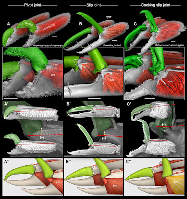
Figure 1. MicroCT Images, Torque Moment Arms, and Schematic Illustrations of Three Shrimp Claw-Joint Types When Closed and Fully Open.
(A) Pivot joint: anterior face∗ of right P1 in a basally branching caridean shrimp. (B) Simple slip joint (no torque reversal or power amplification): anterior face∗ of right P2 in an “intermediate” caridean shrimp. (C) Cocking slip joint (type 1 torque-reversal cocking, most likely power-amplified closing): anterior face∗ of right P1 in a feebly snapping alpheid shrimp. (A’–C’) Overlaid sagittal plane and surface rendering (via micro-computed tomography [microCT]) of claws of all three species showing torque moment-arms (+, –) when closed (upper) and fully opened (lower and background); negative torque (–) indicates that initial contraction of part of the closer muscle causes cocking. (A”–C”) Schematic representation of all three joint types showing loading orientations of opener and closer muscles. (A”) Pivot joint: purely rotational motion of dactyl. (B”) Slip joint: during opening, the dactylar base both rotates and translates (slips) across the propodal ridge (B). (C”) Cocking slip joint: during opening, the dactylar base both rotates and translates—including an abrupt sliding motion into the fully cocked position, where part of the closer muscle (gold) will generate reversed torque (–), and hence energy storage, because it inserts above the fulcrum (white dot).

 White dots show primary rotation axes (A–A”) or fulcrum points (B–B” and C–C”) for dactylar sliding and rotation. Black dots identify a reference point on the dactylar base. White arrows (A–C and A”–C”) show dactylar base trajectories during opening; closing would follow the same trajectories but in reverse. Red arrows (A’–C’) indicate dorsal-most closer-muscle contraction vectors (labeled V1 in Figure 4). Yellow arrows (A’–C’) represent torque moment arms about the fulcrum. Scale bars, 500 μm (A) and 300 μm (B and C). om, opener muscle; cm, closer muscle; (+), positive (counterclockwise) initial torque during claw closing; (–), negative (clockwise) torque during claw cocking generated by the gold-shaded muscle region in (C”). See also Figure SM1 in Methods S1 (joint-type scoring), Figures S1–S4 (microCT images of all claws), Movies S1A and S1B (actual dactyl motion), Movies S2A–S2E (3D model tests), and Table S1 (joint types of all species). ∗See Supplemental Results (Methods S1) for an explanation of claw-face viewing perspectives.


 Tomonari Kaji, Arthur Anker, Christian S. Wirkner and A. Richard Palmer. 2018. Parallel Saltational Evolution of Ultrafast Movements in Snapping Shrimp Claws. Current Biology.  28(1); 106-113.  DOI: 10.1016/j.cub.2017.11.044 

An adaptation 150 million years in the making phy.so/434192683 via @physorg_com

[Ichthyology • 2018] Basic Description and Some Notes on the Evolution of Seven Sympatric Morphs of Dolly Varden Salvelinus malma from the Lake Kronotskoe Basin (Russia, Kamchatka)

$
0
0

Sympatric morphs of Lake Kronotskoe charrs.
 W, white; L, longhead; N, nosed; S, smallmouth; B, bigmouth

Markevich, Esin & Anisimova, 2018.
 DOI:  10.1002/ece3.3806  

Abstract
The study examines the basic morphological and ecological features of Dolly Varden from Lake Kronotskoe (Russia, Kamchatka). Seven valid morphs different in head proportions, feeding, timing, and place of spawning have been determined in this ecosystem. The basic morphometric characteristics clearly separate Lake Kronotskoe morphs from each other, as well as from its potential ancestor (Dolly Varden). According to CVA analysis, the most notable morphological characteristics determining the mouth position are the length of a lower jaw and rostrum. Furthermore, five of seven morphs inhabit different depth zones of the lake and feed on different food resources. Our data suggest that reproductive isolation may be maintained by temporal/spatial isolation for two morphs with lacustrine spawning, and by spatial isolation only for the rest of the morphs with riverine spawning. The sympatric diversity of the Lake Kronotskoe charrs is exceptionally wide, and there are no other examples for seven sympatric morphs of genus Salvelinus to coexist within a single ecosystem. This study puts forward a three-step hypothetical model of charr divergence in Lake Kronotskoe as a potential ground for future studies.

KEYWORDS: “charr problem”, diversification isolation, functional morphology, spawning, trophic polymorphism


Figure 1 Sympatric morphs of Lake Kronotskoe charrs.
W, white; L, longhead; N, nosed: 1—blunt nosed, 2—sharpnosed, 3—shovelnosed; S, smallmouth; B, bigmouth

Grigorii Markevich, Evgeny Esin and Liudmila Anisimova. 2018. Basic Description and Some Notes on the Evolution of Seven Sympatric Morphs of Dolly Varden Salvelinus malma from the Lake Kronotskoe Basin. Ecology and Evolution. DOI:  10.1002/ece3.3806 


[PaleoOrnithology • 2018] A Flattened Enantiornithine in mid-Cretaceous Burmese Amber: Morphology and Preservation

$
0
0

 young Cretaceous bird trapped in tree resin

 Xing, O'Connor, McKellar, et al., 2018.

Illustration: Cheung Chung Tat

Abstract
Cretaceous amber from Myanmar (∼99 Ma Burmese amber) has become a valuable supplement to the traditional skeletal record of small theropod dinosaurs preserved in sedimentary rocks, particularly for coelurosaurs and enantiornithines. The specimens recovered from this deposit preserve skeletal material and soft tissues in unmatched detail. This provides opportunities to study three-dimensional preservation of soft tissues, microstructure, and pigmentation patterns that are seldom available elsewhere in the fossil record. Ultimately, this line of research provides insights into life stages that are difficult to preserve, the ecology and appearance of the groups involved, and the evolutionary-development of structures such as feathers. Here we describe the most recent discovery from Burmese amber, an articulated skeleton of an enantiornithine bird. This individual has been sectioned along the coronal plane, providing a unique view inside multiple body regions. Osteological observations and plumage patterns support placement within the Enantiornithes, and suggest that the animal may have been a juvenile at the time of death. The specimen has a complex taphonomic history that includes exposure at the surface of a resin flow prior to encapsulation, and may include scavenging by some of the insects trapped within the same amber piece. The chemical composition observed along surface exposures and shallowly buried regions of the body indicate that the specimen has not undergone significant exchange with its surroundings. High iron concentrations are present in regions that preserve soft tissues as carbon films, and calcium distribution corresponds to regions where bones breach the surface of the amber.

Keywords: Enantiornithes; Juvenile; Osteology; Soft tissue preservation


An illustration shows the young Cretaceous bird trapped in tree resin, which would eventually fossilize into amber.
Illustration: Cheung Chung Tat


Conclusions
Decay and interactions with the surrounding resin have obscured many of the features of DIP-V-15102, but X-ray µCT is able to provide some osteological information, and the coronal section yields an interesting picture of preservation. The balance of osteological evidence preserved in DIP-V-15102 points towards a source within Enantiornithes. The plumage preserved is also consistent with this placement. The remains are compacted into an amber thickness of approximately 7 mm, but they provide a better sense of how a relatively complete and moist corpse behaves upon entering this preservational setting. Hopefully, this specimen provides a better search image for future discoveries, improving recovery rates and reducing losses due to specimen preparation.


Lida Xing, Jingmai K. O'Connor, Ryan C. McKellar, Luis M. Chiappe, Ming Bai, Kuowei Tseng, Jie Zhang, Haidong Yang, Jun Fang and Gang Li. 2018. A Flattened Enantiornithine in mid-Cretaceous Burmese Amber: Morphology and Preservation. Science Bulletin. In Press.   DOI: 10.1016/j.scib.2018.01.019

 Dinosaur-Era Bird Found Trapped in Amber  on.natgeo.com/2EyVkep via @NatGeo


[Invertebrate • 2018] Astrophiura caroleae • A New Species of the Remarkable Brittle Star Genus Astrophiura (Echinodermata: Ophiuroidea) from the western Atlantic Ocean

$
0
0

Astrophiura caroleae  Pawson, 2018


Abstract

Astrophiura caroleae, new species, is described from off Curacao in the southern Caribbean, and from the western Gulf of Mexico, in depths of 244 to 434 meters. This new species, the first in the genus Astrophiura to be described from the Atlantic Ocean, has a distinctive combination of characters, including regularly arranged primary plates, large radial shields whose radial edges are in contact for their entire visible length, and prominent tubercles on central and radial plates. The mottled reddish coloration of the dorsal surface of this species usually contrasts with the color of the substratum, rendering it readily visible in situ, despite its disc diameter of less than 10 mm. Like its congeners, A. caroleae is gonochoric, the gonads of females containing conspicuous masses of bright orange eggs that are approximately 165 µm in diameter. DNA Barcoding data are provided for this new species, these are the first for Astrophiura.

Keywords: Echinodermata, Ophiuroidea


Class Ophiuroidea
Superorder Euryophiurida O’Hara et al., 2017

Order Ophiurida sensu O’Hara et al., 2017
Family Astrophiuridae Sladen, 1879

Diagnosis. Ophiurids in which enlarged proximal lateral arm plates form the margin of the disc. (Partly after O’Hara, 2017).

Remarks. The paedomorphic (Stöhr and Martynov, 2016) ophiuroid genus Astrophiura has long been placed in the family Ophiuridae. Hugall et al. (2015) noted that Astrophiura is related to Ophiomisidium and Ophiophycis. Then O’Hara et al. (2017) followed the recommendation of Vadon (1990) and formally resurrected the family Astrophiuridae, noting in their Table 4 (p. 422) that the family contains 22 species (presumably Ophiomisidium with eight species; Ophiophycis, seven species; and Astrophiura, approximately seven species; data derived from the World Ophiuroidea Database—Stöhr et al. 2017).

Astrophiura caroleae new species; holotype (USNM 1463102) disc diameter 8.5 mm, recently dead, photographed in laboratory, Note conspicuous female gonads and free arms.


Image: Darryl Felder

Astrophiura caroleae new species

FIGURE 1. A, holotype (USNM 1463102), disc diameter 8.5 mm, in dorsal view (image by A. Nonaka); B, holotype, ventral view (image by A. Nonaka); C, holotype with major dorsal plates labeled; D, paratype 1 (USNM 1463103), disc diameter 7.0 mm, dorsal view; E, detail of holotype showing a pair of radial shields; F, holotype, showing adoral plates, oral plates with oral papillae, and thin plates covering oral interradii.
Abbreviations: AP, adoral plate; B, basal plate; C, central plate; DAP, dorsal arm plate; IB, infrabasal plate; IR, interradial plate; LAP, lateral arm plate; OIP, oral interradial plates; OP, oral plate; R radial plate; RS radial shield. 

FIGURE 2. A, holotype (USNM 1463102) disc diameter 8.5 mm, recently dead, photographed in laboratory (Image: Darryl Felder), Note conspicuous female gonads and free arms. B, holotype in situ on discarded beer bottle; C enlarged image of holotype in situ; D, non-type specimen, in laboratory, disc diameter ca. 8.0 mm, showing color pattern.

Astrophiura caroleae new species

Diagnosis. Astrophiura with disc regularly pentagonal, up to 10 mm in diameter, interradial margins convex. Free portions of arms slightly shorter than disc diameter. In dorsal disc, elongated diamond-shaped radial areas higher than interradial areas. Conspicuous ossicles in dorsal disc include central plate, infrabasals, basals, and radials, three or four interradial plates, the distalmost approximately triangular with a sharp distal point, and large radial shields, pairs of which are contiguous for their entire length. Most plates smooth, but central and radial plates have a single prominent central tubercle, and each radial shield has a more or less conspicuous prominence near the distal edge. Proximal dorsal arm plates on the disc are quadrangular, wider than long. Six or seven oral papillae on each jaw. Adoral plates elongate, visible portions at least 6 times as long as wide, the proximal ends of each pair abutting more or less in line with the apex of each jaw. Color in life variegated brownish to reddish with whitish patches, the tubercles on dorsal plates usually lighter in color than surrounding areas.

 Etymology. It is a pleasure to name this species for Dr. Carole Baldwin, Chair of the Vertebrate Zoology Department, National Museum of Natural History, Smithsonian Institution. Dr. Baldwin founded the Smithsonian’s Deep Reef Observation Project (DROP) in 2011 and she has inspired its steady growth in addition to conducting her own excellent research on fishes.

Preferred Habitats: It is well-known that Astrophiura prefers a hard substratum; individuals can be found attached to dead mollusk shells (Fujita and Hendler, 2001), and rocks (Ziesenhenne, 1951; Matsumoto, 1917). A. caroleae was first observed attached to a discarded Heineken beer bottle, where its reddish coloration contrasted sharply with the green of the bottle. Other specimens were collected from miscellaneous bottles near the type locality

Distribution: This species is known from off Curacao in the southern Caribbean, and also from off Quintana Roo, Mexico, in depths of 244 to 434 meters.


David L. Pawson. 2018. A New Species of the Remarkable Brittle Star genus Astrophiura (Echinodermata: Ophiuroidea) from the western Atlantic Ocean. Zootaxa. 4378(2); 257–264. DOI: 10.11646/zootaxa.4378.2.4


[Botany • 2018] Genomics of the Origin and Evolution of Citrus

$
0
0

Proposed origin of Citrus and ancient dispersal routes. Arrows suggest plausible migration directions of the ancestral citrus species from the centre of origin—the triangle formed by northeastern India, northern Myanmar and northwestern Yunnan. The proposal is compatible with citrus biogeography, phylogenetic relationships, the inferred timing of diversification and the paleogeography of the region, especially the geological history of Wallacea and Japan. The red star marks the fossil location of C. linczangensis. Citrus fruit images in c and d are not drawn to scale.

Wu, Terol, Ibanez, et al., 2018. 

Abstract
The genus Citrus, comprising some of the most widely cultivated fruit crops worldwide, includes an uncertain number of species. Here we describe ten natural citrus species, using genomic, phylogenetic and biogeographic analyses of 60 accessions representing diverse citrus germ plasms, and propose that citrus diversified during the late Miocene epoch through a rapid southeast Asian radiation that correlates with a marked weakening of the monsoons. A second radiation enabled by migration across the Wallace line gave rise to the Australian limes in the early Pliocene epoch. Further identification and analyses of hybrids and admixed genomes provides insights into the genealogy of major commercial cultivars of citrus. Among mandarins and sweet orange, we find an extensive network of relatedness that illuminates the domestication of these groups. Widespread pummelo admixture among these mandarins and its correlation with fruit size and acidity suggests a plausible role of pummelo introgression in the selection of palatable mandarins. This work provides a new evolutionary framework for the genus Citrus.

Figure 1: Genetic structure, heterozygosity and phylogeny of Citrus species.  a, Principal coordinate analysis of 58 citrus accessions based on pairwise nuclear genome distances and metric multidimensional scaling. The first two axes separate the three main citrus groups (citrons, pummelos and mandarins) with interspecific hybrids (oranges, grapefruit, lemon and limes) situated at intermediate positions relative to their parental genotypes. b, Violin plots of the heterozygosity distribution in 58 citrus accessions, representing 10 taxonomic groups as well as 2 related genera, Poncirus (Poncirus trifoliata, also known as Citrus trifoliata) and Chinese box orange (Severinia). White dot, median; bar limits, upper and lower quartiles; whiskers, 1.5× interquartile range. The bimodal separation of intraspecies (light blue) and interspecies (light pink) genetic diversity is manifested among the admixed mandarins and across different genotypes including interspecific hybrids. Three-letter codes are listed in parenthesis with additional descriptions in Supplementary Table 2. c, Chronogram of citrus speciation. Two distinct and temporally well-separated phases of species radiation are apparent, with the southeast Asian citrus radiation followed by the Australian citrus diversification. Age calibration is based on the citrus fossil C. linczangensis16 from the Late Miocene (denoted by a filled red circle). The 95% confidence intervals are derived from 200 bootstraps. Bayesian posterior probability is 1.0 for all nodes. d, Proposed origin of citrus and ancient dispersal routes. Arrows suggest plausible migration directions of the ancestral citrus species from the centre of origin—the triangle formed by northeastern India, northern Myanmar and northwestern Yunnan. The proposal is compatible with citrus biogeography, phylogenetic relationships, the inferred timing of diversification and the paleogeography of the region, especially the geological history of Wallacea and Japan. The red star marks the fossil location of C. linczangensis. Citrus fruit images in c and d are not drawn to scale.

Guohong Albert Wu, Javier Terol, Victoria Ibanez, Antonio López-García, Estela Pérez-Román, Carles Borredá, Concha Domingo, Francisco R. Tadeo, Jose Carbonell-Caballero, Roberto Alonso, Franck Curk, Dongliang Du, Patrick Ollitrault, Mikeal L. Roose, Joaquin Dopazo, Frederick G. Gmitter, Daniel S. Rokhsar and Manuel Talon. 2018. Genomics of the Origin and Evolution of Citrus.  Nature.   DOI: 10.1038/nature25447


The Citrus Family Tree  on.natgeo.com/2AQGWMo   @NatGeoMag


[Paleontology • 2018] Titanosaur Dinosaurs from the Upper Cretaceous of La Rioja Province, NW Argentina

$
0
0

Titanosaur dinosaurs were found in two Upper Cretaceous units of La Rioja, Argentina.

in Hechenleitner, Fiorelli, Martinelli & Grellet-Tinner, 2018.

Highlights
• Titanosaur dinosaurs were found in two Upper Cretaceous units of La Rioja, Argentina.
• Most of the specimens are interpreted as non-Saltasaurinae lithostrotians.
• Few bones (caudal vertebrae) show features consistent with pathology.
• Differences in caudal vertebrae suggest the presence of at least two taxa.

Abstract
Our current knowledge of Argentinean titanosaurs outside Patagonia has been effectively limited to the province of Salta (e.g., Saltasaurus). However, several skeletal remains of titanosaurian sauropod dinosaurs have been discovered in the Cretaceous strata of La Rioja Province, northwest Argentina. Herein we describe new titanosaur material from two Upper Cretaceous localities from La Rioja. The first record corresponds to two caudal vertebrae found in the exposures of the Ciénaga del Río Huaco Formation, in the Quebrada de Santo Domingo locality. The largest sample, which includes fragmentary postcranial material of five individuals, was recovered from the Los Llanos Formation, in the Tama locality. The comparative analyses conducted here suggest that most of the specimens should be referred to as Lithostrotia non Saltasaurinae. However, differences between the middle caudal vertebrae from Tama and Quebrada de Santo Domingo suggest that the taxa at each locality are distinct.

Keywords: Los Llanos Formation; Cienaga del Rio Huaco Formation; Tama; Quebrada de Santo Domingo; Sauropods


Systematic paleontology

Saurischia Seeley, 1888
Sauropodomorpha Huene, 1932

Sauropoda Marsh, 1878
Titanosauria Bonaparte and Coria, 1993
Lithostrotia Upchurch, Barrett and Dodson, 2004

Lithostrotia gen. et sp. indet. 1

Material. CRILAR-Pv 518a-i, nine partial caudal vertebrae.

.....


Conclusions: 
The sauropod specimens described from La Rioja province herein are all referred to Titanosauria. CRILAR-Pv 518 (nine caudal vertebrae) can be identified as a non-saltasaurine lithostrotian, as it presents strongly procoelous centra in its anterior and middle caudal vertebrae, a synapomorphy of Lithostrotia (Upchurch et al., 2004). In addition the centra are almost as high as wide, instead of dorsoventrally compressed as is typical in Saltasaurinae.

The information provided by the few appendicular elements of the specimen CRILAR-Pv 509 suggests that it could be also considered as a non-saltasaurine lithostrotian, as it shows remarkable similarities to Aeolosaurus, Uberabatitan and Bonitasaura, such as the pubis being elongate and only slightly recurved, differing from Saltasaurinae, and the condylar surfaces of the distal end of the femur are perpendicular to its major axis rather than “beveled”.

The specimens CRILAR-Pv 510 and 511 are fragmentary and not particularly diagnostic. However, the procoely of the small centrum of the caudal vertebra CRILAR-Pv 510, as well as its proportions, suggest titanosaur affinities. Considering the taxa represented in the Los Llanos Formation, the ilium CRILAR-Pv 511 has a relative size and a series of characters (such as the lateral expansion preacetabular crest, and the extensive and continuous union between the pubic peduncle and the preacetabular crest), which could only be consistent with the morphology of the Titanosauria.

The poor preservation of the specimen CRILAR-Pv 525 obscures a clear diagnosis but the evidence suggests that its anomalous structures have a pathological origin. Both the presence of bony overgrowth and draining channels (subperiosteal pits) suggest that the origin of this condition was a response to an infection. This constitutes an additional paleopathological record for titanosaurs, which were unreported until recently.

With respect to their diversity, the fragmentary condition of all the specimens of Tama prevents comparison between individuals. Therefore, there is no reason presently to infer the occurrence of more than one taxon in this locality. However, the strong curvature (in lateral view) of the ventral edge of the vertebral centra of Quebrada de Santo Domingo clearly contrasts with the almost flat condition observed in the Tama specimen, suggesting the presence of distinct taxa in each locality.+


E. Martín Hechenleitner, Lucas E. Fiorelli, Agustín G. Martinelli and Gerald Grellet-Tinner. 2018. Titanosaur Dinosaurs from the Upper Cretaceous of La Rioja Province, NW Argentina. Cretaceous Research. 85; 42–59.  DOI: 10.1016/j.cretres.2018.01.006


[Hexapoda • 2018] Rambutanura hunanensis • First Record of the Tropical Genus Rambutanura (Collembola: Neanuridae: Neanurinae) in Palearctic, with Description of A New Species from China

$
0
0

Rambutanura hunanensis  Jiang & Dong 

in Dong, Yang, Zhao, Wang & Jiang, 2018.  

Abstract

Genus Rambutanura Deharveng, 1988 of subfamily Neanurinae is herein recorded and a new species is described from China. A key to the species of the whole world of the genus is provided. In order to determine the new species, a new diagnosis within the genus Rambutanura Deharveng, 1988 is proposed. The new species is distinguished from all known members of the genus by its unique set of morphological characters notably: 2+2 depigmented eyes, mandible quadridentate, presence of 1 additional s-chaeta on each tubercle Di from Th. I to Abd. IV, presence of 2 additional s-chaetae on tubercle Dl from Abd. I to Abd. V, presence of guard chaetae sgd and sgv on AOIII, absence of digitate body tubercles.

Keywords: Collembola, Paleonurini, Rambutanura hunanensis Jiang & Dong sp. nov., taxonomy, key


LIing Dong, Yue-Li Yang, Yu-Yao Zhao, Yuan-Jun Wang and Ji-Gang Jiang. 2018. First Record of the Tropical Genus Rambutanura (Collembola: Neanuridae: Neanurinae) in Palearctic, with Description of A New Species from China. Zootaxa. 4378(3); 377–386.  DOI:  10.11646/zootaxa.4378.3.6


[Entomology • 2018] Dodona formosana • A Distinct Species detected in the Dodona eugenes Species Complex: Clarification of the Taxonomic Status of the Punch Butterfly in Taiwan

$
0
0

Dodona formosana Matsumura, 1919 

in Wu, Lin & Hsu, 2018.
Abstract
The Tailed Punch, Dodona eugenes, is widely distributed in East Asia with seven subspecies currently recognized. However, two of them, namely ssp. formosana and ssp. esakii found in Taiwan, are hard to distinguish from each other due to ambiguous diagnostic characters. In this study, their taxonomic status is clarified by comparing genitalia characters and phylogenetic relationships based on mitochondrial sequences, COI and COII (total 2211 bps). Our results show that there is no reliable feature to separate these two subspecies. Surprisingly we found that Dodona in Taiwan is more closely related to the Orange Punch, D. egeon, than to other subspecies of D. eugenes. Therefore, the following nomenclatural changes are proposed: Dodona eugenes formosana is revised to specific status as Dodona formosanaMatsumura, 1919, stat. rev., and ssp. esakii is sunk to a junior synonym of Dodona formosana syn. n.

Keywords: endemism, male genitalia, Myrsinaceae, wing pattern


Figure 5. Pinned specimens of Dodona formosana and their relatives.
 A D. formosana (spp. formosana) ♂ B D. formosana (spp. formosana) ♀ C D. formosana (spp. esakii) ♂ D D. formosana (spp. esakii) ♀
E D. eu. eugenes♂ F D. eu. venox♂ G D. egeon ♂ H D. egeon
I D. maculosa♂ J D. maculosa ♀ K type of D. formosana (SEHU).

Systematics
Based on the evidence from genitalia morphology and phylogenetic relationships, Dodona from Taiwan should be separated from D. eugenes and regarded as a distinct species. We therefore raised the Dodona butterfly in Taiwan to full species status, Dodona formosana Matsumura 1919, stat. rev., The two previously recognized subspecies, spp. formosana, and spp. esakii, are thus recognized as synonyms (syn. n.).

Dodona formosana Matsumura, 1919, stat. rev.

....

Conclusions
The Dodona butterfly in Taiwan should be regarded as an endemic species, Dodona formosana, distinct from D. eugenes. The present work and a previous study (Callaghan 2009) both point out that Dodona eugenes, widely distributed in East Asia, is not a monophyletic species. Combining evidence from genitalia characters and molecular sequences provides effective clarification on solving species-level problem for species complex containing superficially similar species.


 Li-Wei Wu, Wen-Jie Lin and Yu-Feng Hsu. 2018. A Distinct Species, Dodona formosana, detected in the Dodona eugenes Species Complex: Clarification of the Taxonomic Status of the Punch Butterfly in Taiwan. ZooKeys. 736: 59-77.  DOI: 10.3897/zookeys.736.22062

[Herpetology • 2017] Surveying Europe’s Only Cave-Dwelling Chordate Species, Proteus anguinus, Using Environmental DNA

$
0
0


Abstract
In surveillance of subterranean fauna, especially in the case of rare or elusive aquatic species, traditional techniques used for epigean species are often not feasible. We developed a non-invasive survey method based on environmental DNA (eDNA) to detect the presence of the red-listed cave-dwelling amphibian, Proteusanguinus, in the caves of the Dinaric Karst. We tested the method in fifteen caves in Croatia, from which the species was previously recorded or expected to occur. We successfully confirmed the presence of P. anguinus from ten caves and detected the species for the first time in five others. Using a hierarchical occupancy model we compared the availability and detection probability of eDNA of two water sampling methods, filtration and precipitation. The statistical analysis showed that both availability and detection probability depended on the method and estimates for both probabilities were higher using filter samples than for precipitation samples. Combining reliable field and laboratory methods with robust statistical modeling will give the best estimates of species occurrence.

Proteus anguinus
 Drawing: Marija Crnčec. 

Fig 1. Sampling locations of 16 caves in Croatia.
   Insert (right) shows location of Croatia in Europe (black). Dotted area shows approximate range of P. anguinus. Drawing of P. anguinus is courtesy of Marija Crnčec.

Judit Vörös, Orsolya Márton, Benedikt R. Schmidt, Júlia Tünde Gál and Dušan Jelić. 2017. Surveying Europe’s Only Cave-Dwelling Chordate Species (Proteusanguinus) Using Environmental DNA. PLoS ONE. 12(1): e0170945.  DOI: 10.1371/journal.pone.0170945


Viewing all 10274 articles
Browse latest View live


<script src="https://jsc.adskeeper.com/r/s/rssing.com.1596347.js" async> </script>